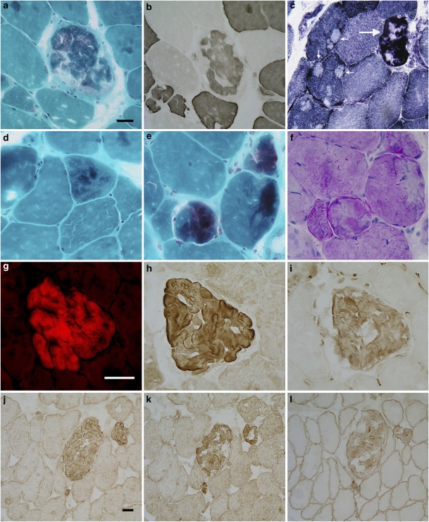
Figure 2.
Histochemical and immunohistochemical findings in a patient with filaminopathy. (a) A large fiber containing a hyaline inclusion. (b) ATPase and (c) oxidative activities are partially reduced in the abnormal fiber area. Some adjacent fibers show several core-like lesions (arrows in c). Other types of lesions: (d) a thin, vermiform inclusion, (e) three muscle fibers containing demarcated abnormal areas, and (f) displaying some PAS positivity. Serial consecutive sections show an inclusion displaying congophilia (g), filamin C (h), ubiquitin (i), desmin (j), myotilin (k), and dystrophin (l) immunoreactivity. (a, d, and e) modified trichrome; (b) ATPase 4.35; (c) NADH; (f) PAS; (g) Congo Red; (h) filamin C; (i) ubiquitin; (j) desmin; (k) myotilin; and (l) dystrophin staining. Bar in a=40 μm (refers to a through f); bar in g=50 μm (refers to g through i); bar in j=25 μm (refers to j through l).

