Figure 2.
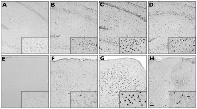
Talampanel protects against later increases in susceptibility to seizure-induced neuronal injury. At P30–31 (20 days after the P10 hypoxic seizures), kainate was administered i.p. (10 mg/kg) and rats were killed 48 h after seizure induction (P33–34). DNA fragmentation is shown in area CA1 and CA3 of hippocampus (A–D) and the basolateral amygdala (E–H). Naive controls (no hypoxia or kainate-induced seizure) show no appreciable staining in hippocampus (A) or amygdala (E). Control litter mates that did not undergo hypoxia at P10 but did undergo kainate- induced seizures on P30–P31 show less DNA fragmentation in hippocampus (B) and amygdala (F) compared to animals that underwent both hypoxia at P10 and kainate-induced seizures at P30–31 in hippocampus (C) and amygdala (G). Talampanel pretreatment before P10 hypoxia reduced later kainate-induced cell death in hippocampus (D) and amygdala (H). Box insets are higher-magnification views of CA1 hippocampal subfields (A–D) and basolateral amygdala (E–H). Scale bars = 50 μm.

