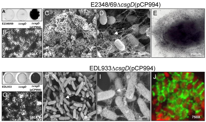
Fig. 3.
Role of curli in biofilm formation. (A) and (F) Crystal violet uptake by biofilms produced by wild-type, ΔcsgD, and ΔcsgD(pCP994) strains on glass coverslips. (B) and (G) Light microscopy micrographs AEEC ΔcsgD(pCP994) biofilms. (C), (D), (H) and (I) SEM micrographs of ΔcsgD(pCP994) biofilms at low and high magnification indicated by scale bars. (E) Immunogold labelling of curli on E2348/69ΔcsgD(pCP994) obtained from the biofilm assay. (J) Detection of curli (green peritrichous fibers) on EDL933ΔcsgD(pCP994) (red bacteria) by immunofluorescence. Biofilms were obtained on a glass substratum after growth of the bacteria at 37°C in T-medium for 24 h. Light microscopy micrographs are at 170X or 750X as indicated.

