
Figure 1.
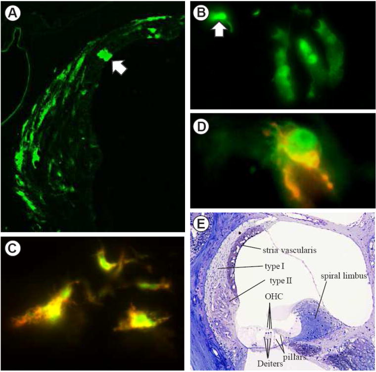
Fluorescent micrographs of GFP in NF-κB reporter mice (A-D). A. Spiral ligament in the lower basal turn of a mouse exposed to 100 dB noise for 2 hours and sacrificed 24 hours post-exposure. Most of the fluorescent cells are type I fibrocytes. The arrow indicates a rare fluorescent marginal cell of the stria vascularis. For orientation, see panel E. B. GFP fluorescent Deiters cells in a control mouse that was not exposed to noise or inflammatory stress. This is a rare case in which 3 adjacent Deiters cells were fluorescent. The arrow indicates the head of an outer pillar cell. C. Co-localization of green GFP fluorescence and red fluorescence reporting anti-GFP. D. A type II fibrocyte in a reporter mouse that was injected with LPS 24 hours prior to sacrifice showing green GFP fluorescence that was largely in different cellular compartments from the red immunostaining for GFP. E. A semithin section stained with osmium and toluidine blue showing the major structures of the cochlear duct. Outer hair cells (OHC), which are sensory cells, rest upon Dieters cells. The lateral wall of the duct is formed by the spiral ligament. Within the ligament the characteristic locations of type I and type II fibrocytes are indicated. The spiral limbus is situated toward the center of the cochlea.