
Figure 2.
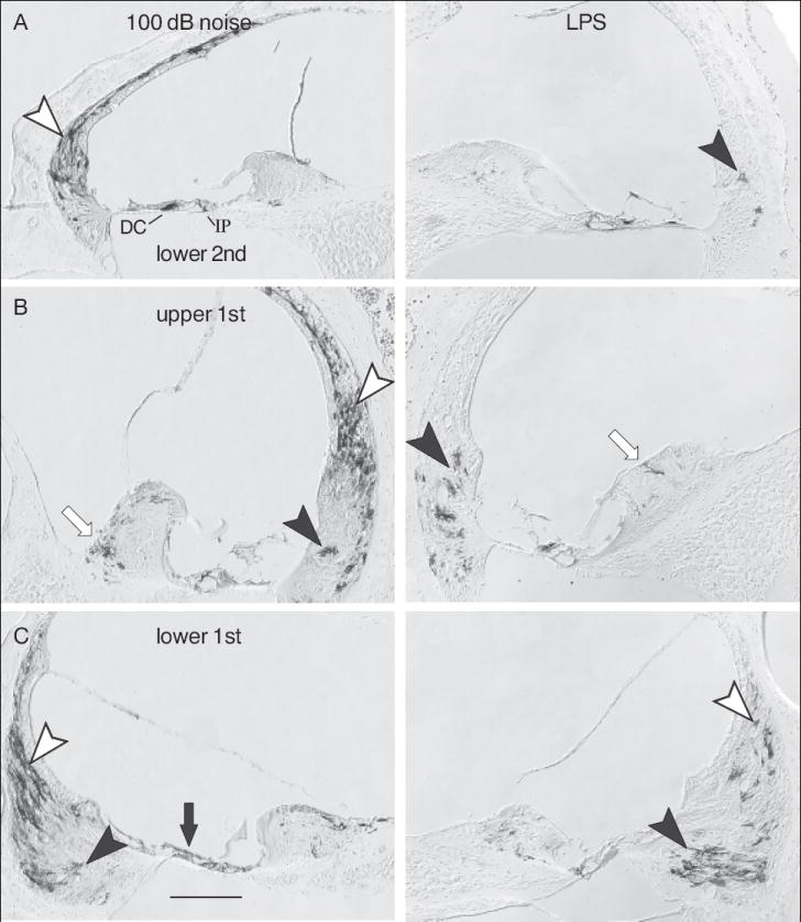
Immunostaining for GFP positive cells in GRP reporter mice following noise exposure and LPS. Each column shows the basal-most three half turns of an ear from a mouse that was exposed to a 100 dB noise (left column) or to intraperitoneal LPS injection (right column). Open arrowheads indicate type I fibrocytes, closed arrowheads indicate type II fibrocytes. Open arrows indicate fibrocytes in the spiral limbus. Closed arrows indicate epithelial cells on the basilar membrane. In panel A, IP indicates inner pillar cell; DC indicates Deiters cell. Calibration bar in C is 100 μm.