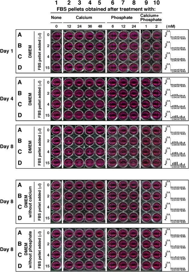
Figure 1. Calcium and phosphate added to serum produce particle-seeding pellets.
Serum pellets were obtained by adding calcium and/or phosphate to FBS to the amounts indicated at the top and incubated overnight at room temperature. Treatment of serum was divided into 4 groups of ions added: no exogenous ions (column 1, “None”); CaCl2 (columns 2–5, referred as “Calcium,” 12–48 mM); Na2HPO4 (columns 6–8, “Phosphate,” 6–24 mM); and a combination of both CaCl2 and Na2HPO4 (columns 9 and 10, “Calcium+Phosphate,” at either 1 mM or 2 mM each). After overnight incubation, the pellets were obtained and processed as described in the Materials and Methods . The resuspended pellets were inoculated into either serum-free DMEM (rows A–D, top 3 panels), calcium-free DMEM (rows A–D, 4th panel from top), or phosphate-free DMEM (rows A–D, bottom panel) in the same order, as three exact replicas. The amount of resuspended pellet volume inoculated into each well is depicted on the left. Observations were done on days 1, 4, and 8 for the top three panels and on day 8 for the bottom two. All incubations were done at 37°C in cell culture conditions. The wells in column 1 corresponded to inoculation with serum pellets obtained from FBS without any ion treatment, while the wells in row A represented control DMEM, without any pellet inoculation. These controls showed no turbidity by visual inspection. On the other hand, turbidity and the presence of precipitates could be seen in all the other wells of the “Day 4” and “Day 8” panels. The wells of the 4th panel, corresponding to incubation in DMEM without calcium, did not show any precipitate whereas the bottom panel wells (DMEM without phosphate) produced small amounts of precipitates.

