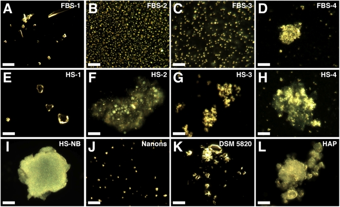
Figure 2. Serum pellets and NB have similar pleomorphic morphologies under dark field microscopy.
Serum pellets were prepared from untreated serum (A and E) or following addition of either 48 mM CaCl2 (B and F), 24 mM Na2HPO4 (C and G), or a combination of both 2 mM CaCl2 and 2 mM Na2HPO4 (D and H) to the indicated serum, followed by incubation at room temperature overnight, centrifugation, and washing steps as described in the Materials and Methods . Low amounts of large heterogeneous particles were noted in the untreated serum pellets (A and E) while the other serum pellets (B–D, F–H) produced smaller and more homogeneous round particles. Such round particles tended to aggregate forming clumps and granular patches (D, F–H). Note that individual granules can be discerned against a background of aggregated material. Similar morphologies were noted in the controls, with HS-NB (I) showing an aggregated diffuse mass in which granules are embedded; “nanons” (J) as dispersed round particles; and DSM 5820 (K) as clumps of highly refringent particles. (L) HAP is shown for comparison. Scale bars: 10 µm.

