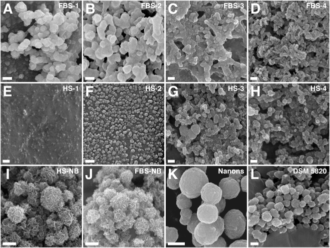
Figure 3. Morphology of serum pellet granulations seen under SEM demonstrates resemblance to NB.
Serum pellets were prepared as in Fig. 2 from untreated serum (A and E) or after addition of either CaCl2 (B and F), Na2HPO4 (C and G), or a combination of both CaCl2 and Na2HPO4 (D and H) to the indicated serum (amounts of ions added as described in Materials and Methods ). Serum pellets primarily harbored round particles in FBS pellets (A–C) and these particles tended to be smaller in HS (E–G). Treatment with both CaCl2 and Na2HPO4 produced particles undergoing different stages of crystallization and film coalescence as well as needle-like projections or spindles (D and H). The morphologies of the serum pellets were similar to the NB controls obtained from either 10% HS (I) or 10% FBS (J) as well as to the NB strains “nanons” (K) and DSM 5820 (L), even though the sizes of the serum pellets particles tended to be smaller than NB. Note that while both “nanons” and DSM 5820 showed smooth surfaces, HS-NB and FBS-NB appeared with rough surfaces containing needle-like crystalline projections. Scale bars: 100 nm (B); 250 nm (C–E, G, H); 300 nm (F, L); 500 nm (I, J); 600 nm (K); 1 µm (A).

