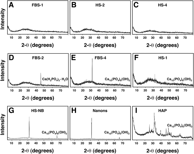
Figure 6. Powder X-ray diffraction spectra of serum granulations demonstrating both amorphous and crystalline patterns.
Serum granulations (pellets) were obtained from either untreated serum (A and F) or following treatments with CaCl2 (B and D) or a combination of CaCl2 and Na2HPO4 (C and E) to the indicated serum, and were dried prior to XRD analysis. Note that the XRD spectra include amorphous patterns (A–C) and peaks corresponding to crystalline compounds of Ca(H2PO4)2·H2O (D) and Ca10(PO4)6(OH)2 (E and F). Crystalline patterns were seen associated with both calcium (D) and calcium phosphate-treated sera (E) as well as untreated serum (F), whereas amorphous patterns were seen not only with untreated serum (A), but also with calcium-treated (B) and calcium phosphate-treated sera (C). XRD spectra corresponding to Ca10(PO4)6(OH)2 were obtained for NB cultured from 10% HS (G) or 10% FBS (H) as well as commercially available HAP used for comparison.

