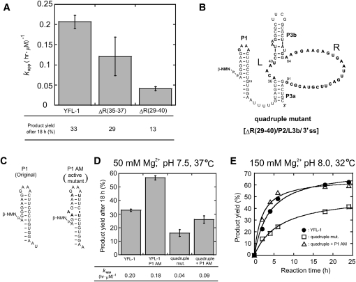
FIGURE 8.
Truncation of the YFL-1 ribozyme. (A) Effects of truncation of the variable region in the catalytic units. ΔR(35–37) and ΔR(29–40) indicate truncated variants, the sequences of which are shown in Figure 1D. (B) Secondary structure of the quadruple mutant with truncations at the tip of P3b, the single-stranded region at the 3′-terminus, P2, and positions R29–40 of the catalytic unit. (C) Secondary structures of (left) the original P1 and (right) its AM variant. (D) Activities of YFL-1, the quadruple mutant, the P1 AM variant, and the double mutant. Reactions were performed with 30 mM Tris-HCl (pH 7.5), 50 mM Mg2+, 200 mM K+ at 37°C. (E) Time courses of the activities of (filled circles) the YFL-1, (open squares) quadruple mutant, and (open triangles) the double mutant derived from the quadruple mutant and the P1 AM variant. Reactions were performed with 30 mM Tris-HCl (pH 8.0), 150 mM Mg2+, 200 mM K+ at 32°C.

