
Figure 4.
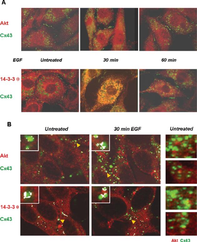
Cx43 co-localizes with Akt and with 14-3-3 θ in vivo. Untreated and EGF-treated (100 ng/mL) Rat-1 cells were immunostained for Cx43 (green) and Akt-3 (red) or 14-3-3 θ (red). (a) Apparent co-localization of the proteins was detected in the EGF-treated cells as shown by the yellow/orange color in the merged images. (b) Confocal microscopy images of the co-stained proteins. Here, the areas of most intense co-localization were highlighted by white pixelation using the confocal microscopy software. The insets in (b) show enlargements of randomly selected Cx43 gap junctional plaques. The arrowheads (yellow) point to the gap junctional plaques enlarged in the insets. Co-localization of Cx43 and Akt is also shown without the white pixilation by the yellow-orange color in the merged images shown on the right-hand side of (b) for untreated cells.