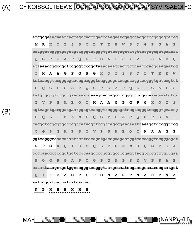
Figure 1.
Topology and sequence of synthetic rlpc gene. (A) Schematic representation and sequence of synthetic LPCcys+. The sequence, derived from the P. yoelii circumsporozoite protein, includes a promiscuous CD4+ T cell epitope, T*-cell (□), an immunodominant B cell epitope from the CSP repeat domain, B-cell (striped bar) and a single CD8+ T cell epitope, CTL (■) arranged in tandem. (B) Amino acid and nucleotide sequences of the synthetic DNA encoding rlpc. A schematic representation of the chimeric protein is included for reference. The amino acid sequence is shown in the single letter code. Four subunits are arranged in tandem interspaced with KAAGPGPG spacers (black boxes in the diagram). The amino acid residues KAA are included downstream of the CD8+ T cell epitope. The carboxyl terminal P. falciparum tag sequence (NANP)3 (underlined) was included for biochemical characterization of the antigenic integrity and to provide optional affinity purification tag. The carboxyl terminal H6 tag sequence (dotted line) was used for protein purification.

