Fig. 5.
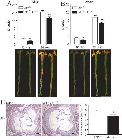
Reduced atherosclerotic lesional development in FP−/− mice. Atherosclerotic lesion area measured by en face staining in male (A) and female (B) Ldlr−/− and Ldlr−/−/FP−/− mice at 12 and 24 weeks on HFD. (C) Aortic root sections (8 μm) from male mice on HFD for 24 weeks were stained by using H&E, with the lesional area calculated. (Scale bar: 100 μm.) Values are presented as mean ± SEM and analyzed by unpaired Student t test, *, P < 0.05 (n = 8–10); **, P < 0.01 (n = 12–16) versus Ldlr−/− control mice.

