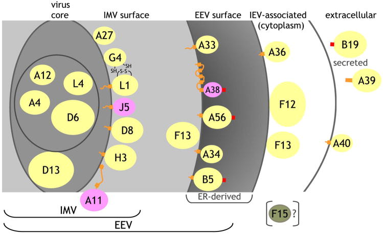
Figure 1. Virus topology of vaccinia proteins used for array.
Based on prior publications and bioinformatics analysis (Table S1), proteins were assigned to the virus core, to the inner membrane of the IMV, to the IMV surface, to the EEV surface, to the outer membrane of the IEV, or as secreted from the host cell. For each protein, the yellow (experimentally demonstrated topology), pink (predicted) or green (F15) ellipse represents the domain synthesized and used in the array where the colored areas are proportional to the spherical size of each protein relative to the others. Not included in the constructed proteins are cytoplasmic domains or short inter-transmembrane regions (orange lines), transmembrane domains (orange blocks), or signal peptides (red blocks). A11 partitions predominantly in the cell but some may associate with the IMV, where the two indicated predicted transmembrane regions do not insert in the IMV membrane. The topology of A38 places it as a Type I integral membrane protein facing into the ER but it has not been unequivocally demonstrated to be on the EEV surface. A39 is secreted in the Copenhagen strain, but is membrane-bound in the vaccinia WR strain. B19 is secreted from the host cell. F15 is not part of the IMV but its topology is uncertain.

