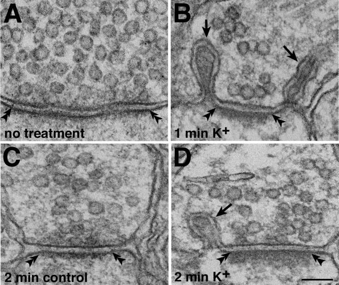
Fig. 2. Depolarization-induced formation of synaptic spinules at excitatory synapses in hippocampal slice cultures.
Synaptic spinules were not found in samples fixed immediately without treatment (A), or in experimental controls (C, 2 min in normal medium). After high K+ depolarization, spinules (arrows in B, D) were found in some synaptic terminal profiles. The postsynaptic densities (PSD, double arrowheads) were relatively thin in A and C compared to those in B and D. Scale bar = 0.1 μm.

