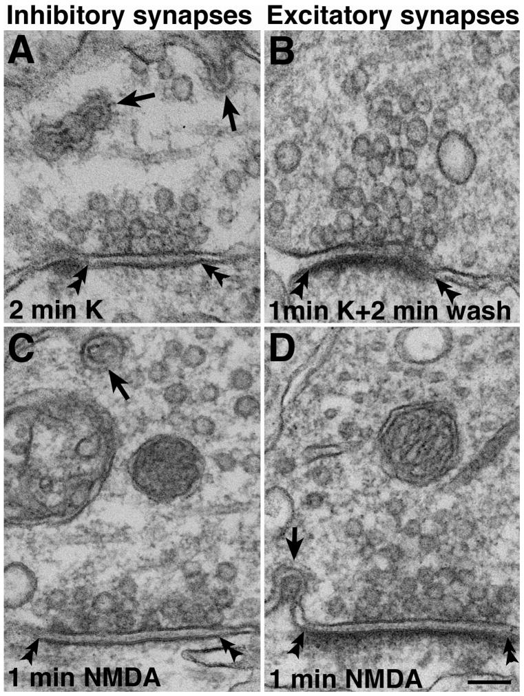
Fig. 4. Synaptic spinule formation and recovery.
(A) - After high K+ depolarization, synaptic spinules (arrows) were also found in inhibitory synapses which lack prominent PSDs (double arrowheads). (B) - Spinules disappeared quickly after cessation of stimulation and a few min of returning to normal medium. The PSD is still thickened in this excitatory synapse. (C) - Spinule (arrow) in an inhibitory synapse after NMDA treatment. (D) - Spinule (arrow) in an excitatory synapse after NMDA treatment. The PSD is visibly thickened. Scale bar = 0.1 μm.

