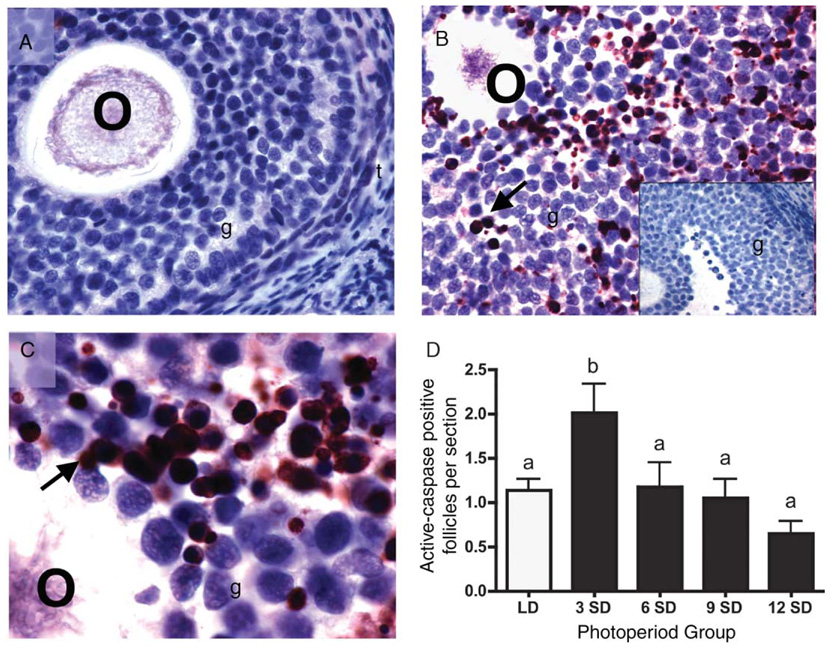
Figure 5.
Representative cross sections from ovaries of Siberian hamsters maintained in LD (A) or 3 weeks of SD (B and C). These sections illustrate the typical lack of active caspase-3 staining in healthy follicles (A), and intense active caspase-3 immunostaining in atretic follicles (B and C). No staining was evident in sections processed without primary antibodies (B inset depicts control atretic follicle). Arrows indicate intense immunolabeling for active caspase-3. O, oocyte; t: thecal layer; g, granulosa cells. × 40 for A, B and B inset; × 100 for C. (D) Quantification of active-caspase-3 positive follicles per ovarian cross section (mean + S.E.M.) in Siberian hamsters housed in long (LD, clear bars; 16L:8D) and short (SD, solid bars; 8L:16D) photoperiods. Groups with different letters are significantly different (P < 0.05).

