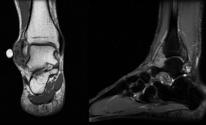
Fig. 1. The magnetic resonance image (coronal and sagittal T1 and fast short-tau inversion-recovery [FSTIR] images) of the patient's right ankle shows an intra-articular well-defined localized mass in the anterolateral ankle joint and sinus tarsi without bony infiltration. The mass has an intermediate density T1 signal and a mildly high FSTIR signal, and it distends the ankle joint locally. On the gradient-echo images, blooming indicated hemorrhage and iron deposition.

An official website of the United States government
Here's how you know
Official websites use .gov
A
.gov website belongs to an official
government organization in the United States.
Secure .gov websites use HTTPS
A lock (
) or https:// means you've safely
connected to the .gov website. Share sensitive
information only on official, secure websites.
