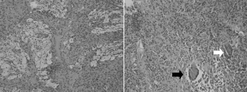
Fig. 2. Characteristic photographs were taken from histologic findings of the bilateral pigmented villonodular synovitis. Left: There is a proliferation of histiocytes, with some (the light coloured cells) showing a lipid-filled cytoplasm (so-called “foamy macrophages”) (hematoxylin and eosin stain, original magnification × 100). Right. A field of histiocytes contains 2 reactive multinucleated histiocytic giant cells. The one on the right (white arrow) has a scattered arrangement of nuclei (a “foreign body” type giant cell), whereas that on the left (black arrow) has clear lipid in its cytoplasm and a wreath-like arrangement of nuclei (a Touton type giant cell) (hematoxylin and eosin stain, original magnification × 200).

An official website of the United States government
Here's how you know
Official websites use .gov
A
.gov website belongs to an official
government organization in the United States.
Secure .gov websites use HTTPS
A lock (
) or https:// means you've safely
connected to the .gov website. Share sensitive
information only on official, secure websites.
