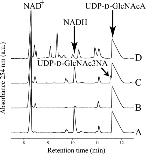
FIGURE 3.
Capillary electropherogram of WbpB, WbpE, and WbpD reactions. A, WbpB enzyme alone produced no product. B, WbpE enzyme alone produced no product. C, WbpB and WbpE incubated together yielded a novel peak that migrated just ahead of UDP-d-GlcNAcA, and this was identified as UDP-d-GlcNAc3NA. D, WbpB, WbpE, and WbpD incubated together do not show the presence of the novel peak, but the electropherogram shows several new peaks caused by the addition of UV-detectable acetyl-CoA (cofactor for WbpD). In all traces, the small peak at ∼11.1 min is a minor contaminant present in the NAD+.

