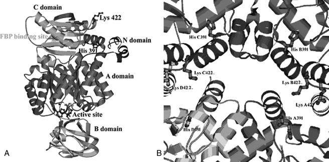
FIGURE 1.
A, ribbon diagram of the overall structure of PK showing the positions of the two mutations, H391Y and K422R, along with the active site and Fru-1,6-P2-binding site. B, intersubunit contact domain of PK. The major amino acid residues and side chains at the tetramer interface region are shown.

