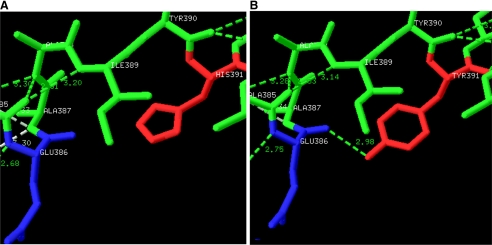
FIGURE 7.
A, the wild-type human PKM2 structure shows His391 (red) of the A-chain and Glu386 (blue) of the C-chain. B, a unique H-bond 2.98 Å in length was predicted between the replaced tyrosine residue (red) and the backbone of Glu386 (blue) connecting the A- and C-domains of the protein. It is hypothesized that formation of a new hydrogen bond connecting the A- and C-domains could restrict the movements along the hinges of the A- and C-chains and cause compromised dynamics in the molecule.

