FIGURE 3.
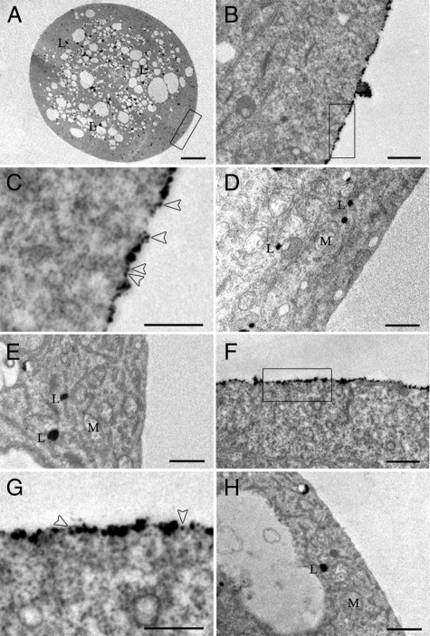
Localization of QD-CaM probe to the outer surface of the plasma membrane of both lily pollen and tobacco BY-2 protoplasts. A–E, transmission electron micrographs of lily pollen protoplasts incubated with QD-CaM probe or unbound QDs. A, low magnification image of a protoplast incubated with QD-CaM probe. B, higher magnification of area highlighted in A. Note that the QD-CaM probe was confined to the outer surface of the protoplast. C, higher magnification of the area highlighted in B. Arrowheads identify single QD-CaM probes bound to the outer surface of the plasma membrane. D and E, lily protoplast incubated with unbound QDs and QD-CaM TR1C probe, respectively. No particles were detected bound to the plasma membrane. F, presence of QD-CaM probe bound to the plasma membrane of a BY-2 protoplast. G, higher magnification of the area highlighted in F. Arrowheads identify single QD-CaM probes bound to the outer surface of the plasma membrane. H, BY-2 protoplast incubated with QD-CaM TR1C probe; no particles were bound to the plasma membrane. Scale bars, A, 10 μm; B–H, 0.5 μm. M, mitochondria; L, lipid.

