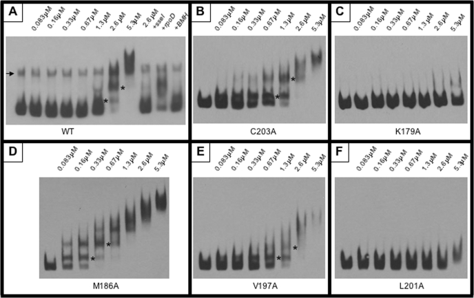
FIGURE 12.
Electrophoretic mobility shift assay. Binding of purified SsrBC and its substituted derivatives to the sseI promoter was determined using an EMSA. Increasing concentrations of protein (as indicated above each lane) were incubated with sseI promoter DNA and separated by PAGE. Protein-DNA complexes migrated more slowly through the gel resulting in a “shift.” EMSAs were performed using identical concentrations of wild-type SsrBC (WT (A)), SsrBC C203A (B), SsrBC K179A (C), SsrBC M186A (D), SsrBC V197A (E), and SsrBC L201A (F). Discrete shifts are indicated by asterisks, and the arrow (in A) indicates the presence of contaminating single-stranded DNA (not present in the other reactions). A, lane 11, indicates that the BMH cross-linked protein is capable of binding to sseI DNA but with reduced affinity (compare with lane 7).

