FIGURE 3.
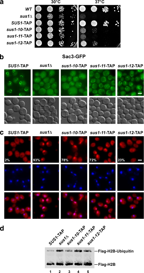
Uncoupling of Sus1 from Sac3 causes TREX-2 mislocalization and mRNA export defects in vivo. a, growth analysis of SUS1-TAP wild-type (WT) and mutant cells on rich medium (YPD) at 30 and 37 °C. Cell density was normalized, and 10-fold serial dilutions were prepared and then plated. Plates were incubated for 2 days. b, subcellular localizations of Sac3-GFP in the indicated wild-type and mutant sus1 strains are shown. Fluorescence microscopic and Nomarski photographs of representative cells grown at 30 °C are shown (scale bar, 2 μm). c, analysis of nuclear mRNA export in the indicated wild-type and sus1 mutant strains. Exponentially growing cells were shifted to 37 °C and grown for 2 more hours before detection of poly(A)+ RNA by fluorescent in situ hybridization with Cy3-labeled oligo(dT) probes. DNA was stained with 4,6-diamidino-2-phenylindole. Numbers indicate the percentage of cells (∼200 cells counted in each case) that exhibit an apparent mRNA export defect. Scale bar, 2 μm. d, analysis of global histone H2B ubiquitin levels. Anti-FLAG-H2B immunoprecipitates derived from SUS1-TAP cells, sus1Δ cells, and the indicated mutants. Recovered proteins were analyzed by SDS-PAGE and Western blotting using an anti-FLAG antibody to detect unmodified FLAG-H2B and ubiquitinated FLAG-H2B.

