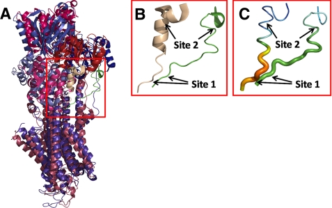
FIGURE 1.
A-M3 linker configuration in E1- and E2-type crystal structures. Crystal structures with Protein Data Bank codes 2zbd (Ca2E1P analog) and 1wpg (E2·Pi analog) are shown aligned. A, overview of structure 2zbd in bluish colors with green A-M3 linker and structure 1wpg in reddish colors with wheat A-M3 linker. B, magnification of the A-M3 linker (corresponding to the red box in A) with arrows indicating site 1, between Glu243 and Gln244, and site 2, between Gly233 and Lys234, in both conformations. The green A-M3 linker to the right is structure 2zbd. The wheat A-M3 linker to the left is structure 1wpg. Note the kinked A3 helix forming part of the latter structure. C, same A-M3 linker structures as in B but with the magnitude of the temperature factor (B-factor) indicated in colors (red > orange > yellow > green > blue) and by tube diameter. Because the two crystal structures selected here as E1- and E2-type representatives have similar crystallographic resolution (2.40 and 2.30 Å, respectively), the differences in temperature factor in specific regions provide direct information about chain flexibility.

