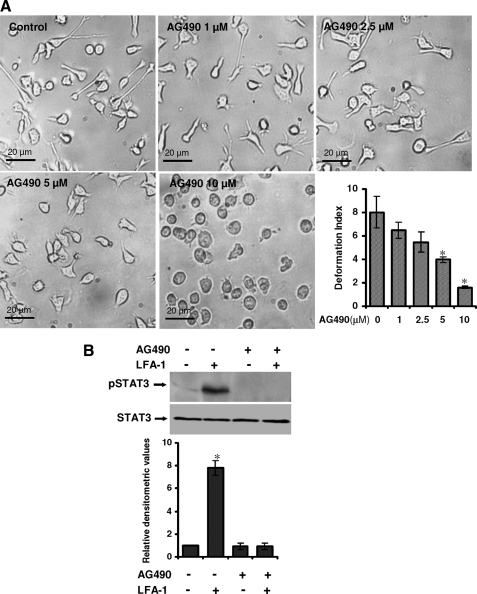
FIGURE 2.
Effect of AG490 on LFA-1-induced locomotory phenotype of T-cells. A, Hut78 cells were pretreated with either vehicle (0.01% (v/v) DMSO; Control) or AG490 (1, 2.5, 5, or 10 μm) for 1 h and incubated on an anti-LFA-1-coated 96-well plate for 4 h. At least 20 microscopic fields were photographed, and a representative image is shown. Dose response migration inhibition by AG490 in Hut78 cells stimulated via immobilized anti-LFA-1 was quantified by measurement of DI and presented. B, untreated or AG490 (10 μm)-treated serum-starved Hut78 cells were stimulated with or without anti-LFA-1 for 10 min and lysed. Cell lysates (20 μg each) were resolved by SDS-PAGE and after Western blotting were probed with anti-phospho-STAT3 (Tyr705; pSTAT3) or anti-STAT3 antibody. Relative densitometric analysis of the individual pSTAT3 band was performed and presented. Data are means ± S.E. of three independent experiments. *, p < 0.05 with respect to control.

