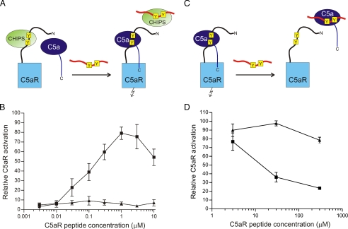
FIGURE 2.
Inhibition of the C5a-induced calcium mobilization. A, schematic representation of the C5a/C5aR7-28S2 inhibition experiment in the presence of CHIPS. The sulfated tyrosines in both the C5aR and C5aR7-28S2 are marked in yellow. Activation of the C5aR is indicated by the flash symbol. B, C5a inhibition experiment in the presence of CHIPS. CHIPS31-121 (10 nm) was preincubated with different concentrations of the C5aR N-terminal peptides C5aR7-28 (triangles) and C5aR7-28S2 (squares) for 30 min at room temperature. Fluo-3-labeled U937/C5aR cells were incubated with these CHIPS31-121/peptide mixtures for 30 s, before stimulation of the cells with 0.3 nm C5a. The increase in fluorescence was measured by flow cytometry. Activation of cells by C5a incubated in the absence of CHIPS and peptide is set to 100%. C, schematic representation of the C5a/C5aR7-28S2 inhibition experiment in the absence of CHIPS. D, C5a inhibition experiment in the absence of CHIPS. C5a (0.3 nm) was preincubated with different concentrations of the C5aR N-terminal peptides C5aR7-28 (triangles) and C5aR7-28S2 (squares) for 30 min at room temperature. These C5a/peptide mixtures were then used for stimulation of Fluo-3-labeled U937/C5aR cells.

