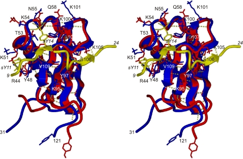
FIGURE 6.
Side-by-side stereo plot displaying the difference between a representative structure of free CHIPS31-121 (red) (PDB ID code 1XEE.pdb) and the corresponding structure of the CHIPS31-121 protein as part of the CHIPS31-121:C5aR7-28S2 complex (blue). Amino acid side chains of CHIPS31-121 that are important for binding C5aR7-28S2 in the core region between residues 9-24, and that have different orientations in the free and unbound form, are displayed in stick representation. The backbone trace of residues 9-24 of C5aR7-28S2 is displayed in yellow with residues sY11 and sY14 in stick representation. The two models mutually overlay with an RMS deviation of 0.089 nm for the well-defined backbone atoms between residues 36-113. The ensemble average backbone RMS deviation is 0.13 ± 0.02 nm.

