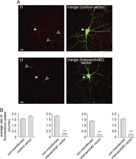
FIGURE 6.
Intersectin KD delays CME of transferrin in hippocampal neurons. Hippocampal neurons were transfected with control vector or intersectin miRNA vectors as indicated. A, transfected cells (green) were identified due to EmGFP expression from the vectors. The neurons were then processed for transferrin (Tf) uptake (red), and images at the end of 30 min of uptake are displayed. Filled arrowheads indicated transfected neurons, and open arrowheads indicate surrounding non-transfected neurons. Movies corresponding to these images are in supplemental Fig. S1. B, the bar graphs indicate the average rate of fluorescent Tf uptake ± S.E. (arbitrary fluorescence units (AU)/min) from a population of neurons coming from at least three separate transfections/cultures. ***, p < 0.001 F-test.

