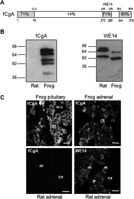
FIGURE 1.
Specificity of the antibody directed against frog CgA. A, scheme depicting the structure of fCgA and showing the high conservation of the terminal regions and the percentages of amino acid identity between frog and human CgA sequences. The highly conserved peptide WE14 and dibasic cleavage sites are also indicated. B, Western blot showing that the antibody developed against fCgA recognized the protein and several processing intermediates in frog but not rat pituitary extracts, whereas an antibody, directed against the WE14 conserved peptide, detected CgA and its processing products in both rat and frog pituitary extracts. C, immunofluorescence analysis of frog pituitary and adrenal glands, and rat adrenal gland using the antibodies against fCgA and WE14. cx, cortex; DL, distal lobe; IL, intermediate lobe; and m, medulla. Scale bars equal 10 μm.

