FIGURE 5.
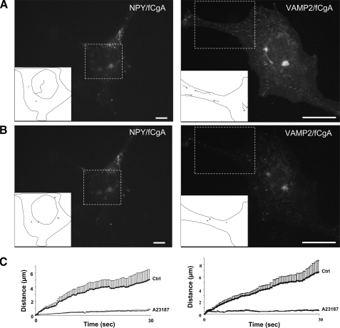
Frog CgA induces the biogenesis of mobile DCG-like structures. A and B, COS-7 cells were cotransfected with full-length fCgA and NPY-Venus or VAMP2-GFP, and imaged via live epifluorescence microscopy before (A) and after (B) treatment with A23187 (1 μm) for 10 min. Small vesicular structures were observed in the cytosol and were tracked during 30 s (supplemental videos S5-S8). Composite images (boxes and insets) containing the tracked vesicles in portions of transfected cells are shown. Few tracked vesicles moved rapidly before A23187 treatment but were essentially immobile after treatment (supplemental videos S5-S8). C, plots of the distance covered from origin against time for the vesicles tracked in control and stimulated conditions.

