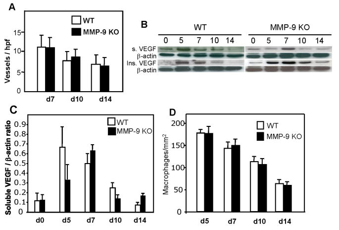
Figure 2. Normal angiogenesis, VEGF levels, and macrophage content in MMP-9-null wounds.
Sections of wounds were stained with anti-PECAM1 antibodies and visualized with the peroxidase reaction. A. Results of vessel quantification for d 7, 10, and 14 wounds. A total of 50 sections per time point per genotype were analyzed. B. Representative Western blots of soluble (top panel) and insoluble VEGF (lower panel) in pooled samples from d 0, 5, 7, 10, and 14 wounds are shown. Immunodetection of β-actin is shown as loading control. The blots were repeated in triplicate with separate extracts and produced similar results. C. Densitometric analysis of western blots showing the ratio of soluble VEGF to β-actin. n = 3. D. Wounds from WT and MMP-9-null mice were stained with the Mac3 antibody and the number of macrophages was quantified from digital images obtained from d 5, 7, 10, and 14 wounds. A total of 50 images from five wounds per time point per genotype were analyzed.

