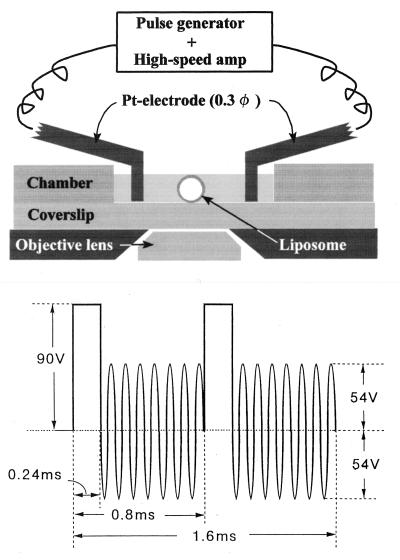
Figure 1.
(Upper) A schematic representation of the electroporation setup. Observations were made in an open chamber. A liposome was placed in the middle of a pair of Pt wire electrodes that were fixed on Plexiglas rods (not shown) for adjustment of the position of the electrodes. (Lower) A shape of the electric pulse used for electroporation.

