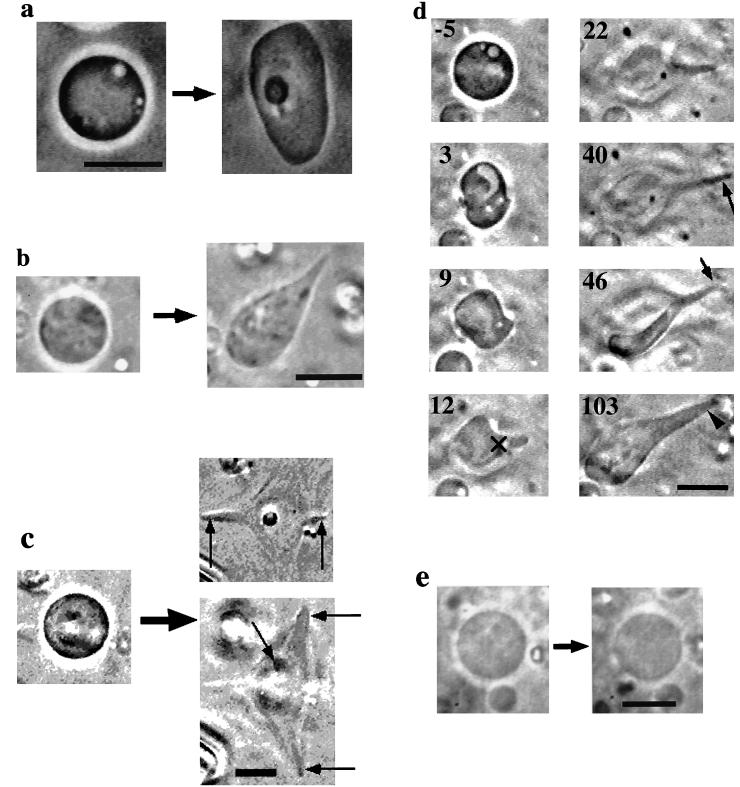
Figure 2.
(a–d) Phase-contrast micrographs of the liposomes containing actin observed after the electroporation. (a) A liposome transforming into an irregular shape; (b and c) Liposomes developing protrusions. In c, the liposome was found to develop five protrusions lying at different focus levels, as indicated with arrows in the panels (Upper Right, lower focus; Lower Right, higher focus). We could record the development of only two of them, indicated with vertical arrows. (d) Process of protrusive growth. Number in each frame represents the time in sec elapsed after the pulse application. The symbol × indicates the reference point used to determine the rate of protrusive formation (see text). (e) Comparison of the shape of a liposome containing 100 μM BSA before and after the electroporation. Bars, 10 μm.

