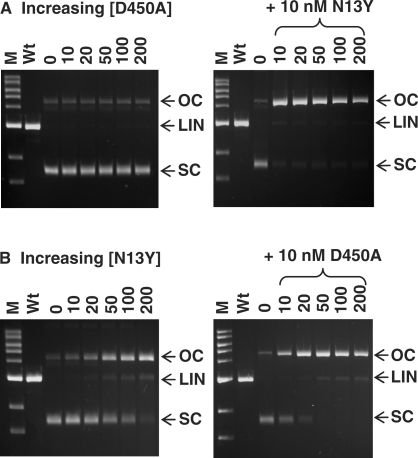
Figure 4.
Specificity of nicking. The reactions, in Buffer 4, contained 5 nM SC pSKFokI (a plasmid with one recognition site for FokI) and FokI protein as indicated below. Reactions were stopped after 1 h at 37°C and the samples analysed by electrophoresis through agarose. The symbols SC, OC and LIN on the right of each gel mark the electrophoretic mobilities of the intact SC DNA, the nicked OC form cut in one strand and the LIN form cut in both strands at one site. The lanes marked M contain 1 kb electrophoresis markers (NEB), and the lanes marked Wt are from equivalent 1 h reactions of 10 nM wt FokI on pSKFokI. (A) Left-hand gel: the reactions contained D450A at the concentrations indicated above each lane (0 → 200 nM). In the right-hand gel, the reactions with 10 → 200 nM D450A also contained 10 nM N13Y. (B) As (A) except that the protein whose concentration was varied was N13Y and that, in the right-hand gel, the samples with varied N13Y also contained 10 nM D450A.

