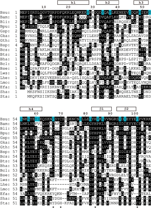
Figure 1.
Alignment of the amino-acid sequence of the N-terminal domain of DnaI from B. subtilis with homologous N-terminal regions of different bacterial helicase loaders. The following sequences are shown (organism, abbreviation, GenBank number): B. subtilis, Bsu, NP_390776; B. amyloliquefaciens, Bam, YP_001422191; B. licheniformis Bli, YP_092605; B. pumilus, Bpu, YP_001487763; Geobacillus sp., Gsp, ZP_02915066; Geobacillus kaustophilus, Gka, YP_148574; Geobacillus thermodenitrificans, Gth, YP_001126735; B. species SG-1, Bsp, ZP_01861306; B. cereus subsp. cytotoxis, Bcs, YP_001376478; B. thuringiensis serovar israelensis, Bts, ZP_00742835; B. halodurans, Bha, NP_244010; B. clausii, Bcl, YP_176196; B. selenitireducens Bse, ZP_02170630; Listeria welshimeri serovar, Lws, YP_849770; Lactobacillus helveticus, Lhe, YP_001577829; L. monocytogenes, Lmo, ZP_02289641; Enterococcus faecium, Efa, ZP_00604060; S. haemolyticus, Sha, YP_253156 and S. aureus, Sta, NP_646444. Residues 1–106 of B. subtilis DnaI correspond to the DnaI-N106 domain for which the 3D structure was determined by NMR spectroscopy. An extended version comprising residues 1–123 (DnaI-N123) was also investigated in the present study. The alignment includes sequences with high homology to B. subtilis DnaI-N106 as identified in a BLAST (47) search. The residue numbers and regular secondary structure elements of B. subtilis DnaI-N106 are indicated at the top. Identically conserved residues are highlighted in black. Residues with <10% side-chain solvent accessibility in the 3D structure are highlighted in blue. Four asterisks at the bottom identify the zinc-binding residues of B. subtilis DnaI.

