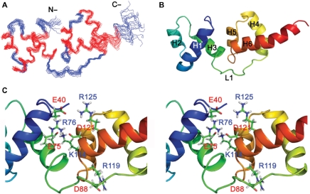
Figure 3.
NMR solution structure of tvMyb135–141. (A) Backbone representation of the ensemble of 20 lowest energy structures. The helical residues are colored in red and others in blue. (B) Secondary structures of the lowest energy structure of tvMyb135–141 displayed in rainbow colors from N-terminus (blue) to C-terminus (red). The first three helices (H1–H3) constitute the R2 motif and the last three helices (H4–H6) form the R3 motif. Two motifs are connected by a long loop (L1). (C) Stereo view of the interface between R2 and R3 motifs. The residues involved in salt bridges between two motifs are shown and labeled.

