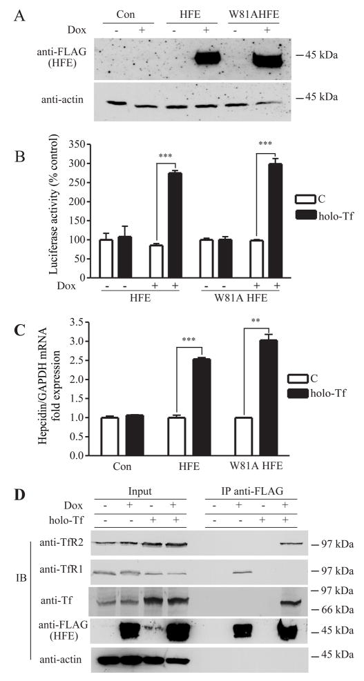
Figure 2. Tf-induced dissociation of HFE from TfR1 results in the association of HFE with TfR2 and stimulates hepcidin promoter activity.
A. Inducible expression of wild-type or W81A mutant HFE in HepG2/tTA HFE and HepG2/tTA W81AHFE cells. Lysates (25 μg) of HepG2/tTA HFE or HepG2/tTA W8AHFE cells, uninduced (−dox) or induced (+dox) to express HFE or W81AHFE, were detected with anti-FLAG antibody. B. HFE is required for Tf to induce hepcidin promoter activity independently of interaction with TfR1. Cells were cotransfected with pLuc-link-HAMP and pCMV-β-gal, treated without (C) or with holo-Tf and analyzed as described in Experimental Procedures. Results are expressed as average ± S.D. of three independent experiments performed in triplicate. C. Hepcidin mRNA expression in HepG2/tTA (Con) HepG2/tTA HFE (HFE) and HepG2/tTA W81AHFE (W81AHFE) cells were treated without (C) or with holo-Tf as described in Experimental Procedures. The expression of hepcidin was measured by qRT-PCR and normalized to GAPDH. The fold of increase in hepcidin mRNA in the dox-induced group was obtained by normalization to uninduced controls. All samples were run in triplicate in three independent experiments. Data are shown as average ± S.D. D. Tf releases HFE from TfR1 to bind to TfR2 in HepG2 cells. Cell lysates (200 μg) from HepG2/tTA HFE treated with or without 25 μM holo-Tf were immunoprecipitated with rabbit anti-FLAG antibodies, and Sepharose-4B/Protein A. Proteins were detected on immunoblots using mouse anti- TfR2, TfR1, FLAG, and actin and goat anti-Tf. The input lanes correspond to 1/5 of the material used for immunoprecipitation. These results were repeated once with similar results. P-values < 0.001 are indicated by *** and < 0.01 by **.

