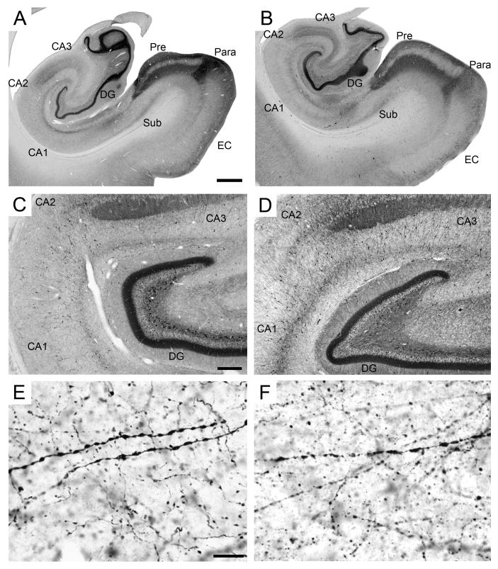
Figure 8.
Calretinin immunoreactivity in the monkey hippocampal formation. A-B: Low magnification photomicrographs of the distribution of calretinin-immunoreactive fibers and cells. A: PM-17-03, perfusion-fixed; B: PM-02-02, immersion-fixed 2 hours postmortem. Scale bar in A: 1 mm, applies to panels A, B. C-D: Intermediate magnification photomicrographs of calretinin-immunoreactivity in the rostral dentate gyrus, CA3 CA2 and CA1 fields of the hippocampus. C: PM-15-03, perfusion-fixed; PM14-03, immersion-fixed 12 hours postmortem. Note the calretinin-positive neurons in the polymorphic layer of the dentate gyrus. Scale bar in C: 300 μm, applies to panels C, D. E-F: High magnification of calretinin-positive fibers in the stratum radiatum of the CA1 field of the hippocampus. E: PM-17-03, perfusion-fixed; F: PM-02-02, immersion-fixed 48 hours postmortem. Note the degradation of cellular labeling in immersion-fixed tissue. Scale bar in E: 15 μm, applies to panels E, F. Abbreviations: see Figure 1.

