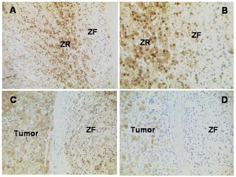
Figure 6.

Immunolocalization of AKR1C3 and CYP19 in an adrenal containing an estrogen-secreting adrenal adenocarcinoma. A. Immunohistochemistry for AKR1C3 showing normal adrenal cortex with higher expression centrally in the lipid-poorer cells of the zona reticularis (ZR) than in the lipid-rich cells of the zona fasciculata (ZF); the adrenal capsule is located at the extreme right of the image (original magnification x20). B. Immunohistochemistry for AKR1C3 showing normal adrenal cortex with higher expression in the lipid-poorer cells of the zona reticularis (centre) than in the lipid-rich cells of the zona fasciculata (right); adrenal capsule to the far right of the image (original magnification x40). C. Immunohistochemistry for AKR1C3 showing expression in the carcinoma cells (left), tumor capsule (centrally) and some of the normal adrenocortical cells (right) (original magnification x20). D. Immunohistochemistry for aromatase, showing expression in many of the carcinoma cells (left) and the normal adrenal cortex to be negative (right) (original magnification x20).
