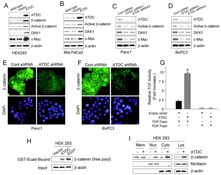
Figure 3. ATDC upregulates β-catenin levels and TCF transcriptional activity.
(A, B) Representative Western blots of wild type, empty vector, and ATDC-transfected HEK 293 (A) and MiaPaCa2 cells (B). Overexpression of ATDC results in upregulation of β-catenin, active β-catenin, and the TCF target genes DKK1 and c-Myc. β-actin was used as a loading control. (C, D) Representative Western blots of Panc1 (C) and BxPC3 (D) cells expressing control shRNA, ATDC shRNA1 or 2. Silencing of ATDC in Panc1 and BxPC3 cells decreases levels of active β-catenin, DKK1 and c-Myc. β-Actin serves as a loading control. (E, F) Photomicrographs of control shRNA and ATDC shRNA1-expressing Panc1 cells (E) and BxPC3 cells (F) immunostained with an anti-β-catenin antibody (green). Cell nuclei were counterstained with DAPI (blue). (G) TCF reporter activity was assessed by using the β-catenin responsive TOPFLASH reporter and the mutant control FOPFLASH reporter in HEK 293 cells stably transfected with empty vector or an ATDC expression vector (mean±SE, *p<0.05 vs empty vector-transfected cells). (H) The GST-E-cadherin (GST-Ecad) fusion protein detects increases in the free pool of β-catenin. HEK 293 cells expressing ATDC or S33Y β-catenin (S33Y) were harvested. Free β-catenin levels were assessed by western blotting of GST-Ecad-bound fractions of cell lysate using a specific anti-β-catenin antibody. β-actin (input) was used as a loading control. (I) Representative blots of β-catenin levels in membrane (Mem), nuclear (Nuc) and cytoplasmic (Cyto) fractions and total lysates (Lys). β-actin (cytoplasmic expression) and fibrillarin (nuclear expression) were used as loading controls.

