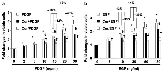
Figure 1.
Curcumin, with different treatment strategies, showed different efficiencies in the inhibition of PDGF- or EGF-stimulated HSC proliferation. Cultured HSCs were divided into three groups. In the first two groups, HSCs were serum-starved in DMEM with 0.5% of FBS for 24 h prior to addition of PDGF (a) or EGF (b) at the indicated doses in the simultaneous presence (Cur + PDGF, or Cur + EGF) or absence (PDGF or EGF) of curcumin (20 μM) for an additional 24 h. In the third group, HSCs were pretreated with curcumin (20 μM) for 24 h in DMEM with 0.5% of FBS prior to the addition of PDGF (a) or EGF (b) at the indicated doses for an additional 24 h (Cur/PDGF, or Cur/EGF). Cell proliferation was determined by colorimetric MTS assays. Results were expressed as fold changes in the number of viable cells, compared with the untreated control (the first columns on the left). Values were expressed as means ± s.d. (n = 3). *P < 0.05 vs the untreated control (the first column on the left). §P < 0.05 vs cells treated with PDGF or EGF only (the left first column in the same group). ‡P < 0.05 vs cells simultaneously treated with curcumin plus PDGF or EGF (Cur + PDGF, or Cur + EGF) (the middle column in the same group).

