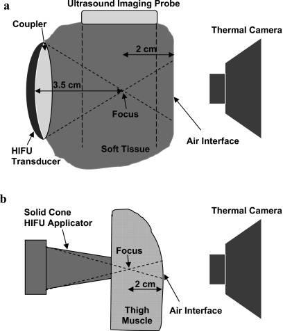
Figure 2.
In-vitro and in-vivo experimental setups. The temperature at the tissue-air interface in the post-focal region was measured using a thermal camera. (a) Treatment of the turkey breast in-vitro was performed using a 3.2 MHz HIFU device (focus at 3.5 cm). HIFU focus was observed using B-mode ultrasound imaging. (b) In-vivo treatment was performed using an intraoperative 5.5 MHz HIFU solid cone applicator (with the focus located at ∼1 cm from the tip of the applicator).

