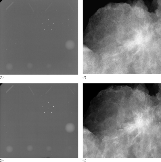
Figure 4.
Conventional (attenuation) images (A, C) and phase contrast images (B, D) of accreditation phantom (top row) and lumpectomy specimen (bottom row). All images were acquired at 40 kVp with same entrance exposure for attenuation and phase contrast images. These images were acquired using prototype systems developed at the University of Oklahoma (Hong Liu), in collaboration with University of Alabama at Birmingham (Xizeng Wu) and University of Iowa (Laurie L. Fajardo). (Courtesy: Hong Liu, University of Oklahoma)

