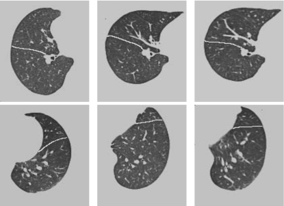
Figure 9.
Slice 151 (top row, the right lung) and 184 (bottom row, the left lung) from a 3D registration experiment. The columns from left to right correspond to the template T, the target S, the deformed template T∘h. Notice a good alignment between the oblique lobar fissures (highlighted in white lines) and between the artery branches, in addition to a good alignment of global shapes after registration.

