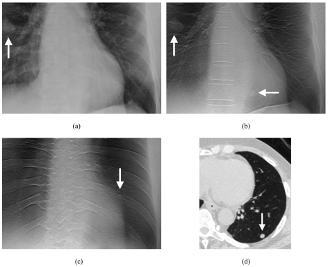
Figure 1.
Images of nodules in one of the human subjects. (a) Coned view of digital PA radiograph shows one clearly visible right lung nodule (arrow). (b) Tomosynthesis image shows the same nodule (vertical arrow) as seen on the PA radiograph in (a). A second nodule (horizontal arrow) is also visible that was not seen in the PA radiograph in (a). (c) Tomosynthesis image at a more posterior level shows an additional left lung nodule (arrow) not seen in the PA radiograph in (a). (d) CT image (lung window) confirms left lower lobe nodule seen in (c).

