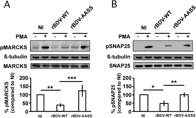
Figure 2. Analysis of PKC signaling in hippocampal neurons infected with the different recombinant viruses.
Total extracts were prepared from non-infected (NI) neurons and neurons infected with the rBDV-WT or rBDV-AASS viruses (21 days post-infection), after stimulation or not with 1 µM PMA for 10 min. Equivalent protein amounts were analyzed by Western blot with specific antibodies for (A) phospho-MARCKS (pMARCKS; PKC site at Ser152/156) and (B) phospho-SNAP-25 (pSNAP-25, PKC site at Ser187). ß-tubulin and total MARCKS and SNAP-25 were used to normalize expression. Bottom graphs show the fluorometric quantification, using the Odyssey imager, of four to ten independent experiments. Quantification results were expressed as percentage of increase relative to the response of NI neurons, which was set at 100%. Values are expressed as mean±s.e.m. *, p<0.05; **, p<0.01; ***, p<0.001 by unpaired t-test.

