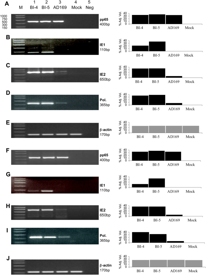
Figure 8. Detection of HCMV ie1, ie2, pol, and pp65 mRNA in human EC.
The viral gene expression was determined by RT-PCR assays at 14 days post infection. The pp65 gene expression served as the control. (A–E) HCMV RNA expression in infected umbilical vein cells (CRL-1730). (F–J) HCMV RNA expression in abdominal aorta cells (CRL-2472). Data show that HCMV clinical isolates, BI-4 and BI-5, expressed viral specific genes and persistently infected EC, whereas HCMV lab strain, AD-169, did not. The RT-PCR products examined by agarose gel are shown on the left panel, and by densitometer tracing are shown on the right panel.

