
Figure 1.
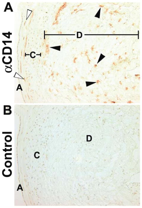
Immunohistochemical localization of macrophages in term extraplacental membranes. (A) Anti-CD14. (B) Isotype matched control. Open arrows mark fetal macrophages between the amnion and chorion membranes. Closed arrows mark decidual macrophages close to the chorion membrane and scattered randomly through the decidua. A, amnion epithelium; C, chorion cytotrophoblast cell layer; D, decidua. Original magnifications, 200×.